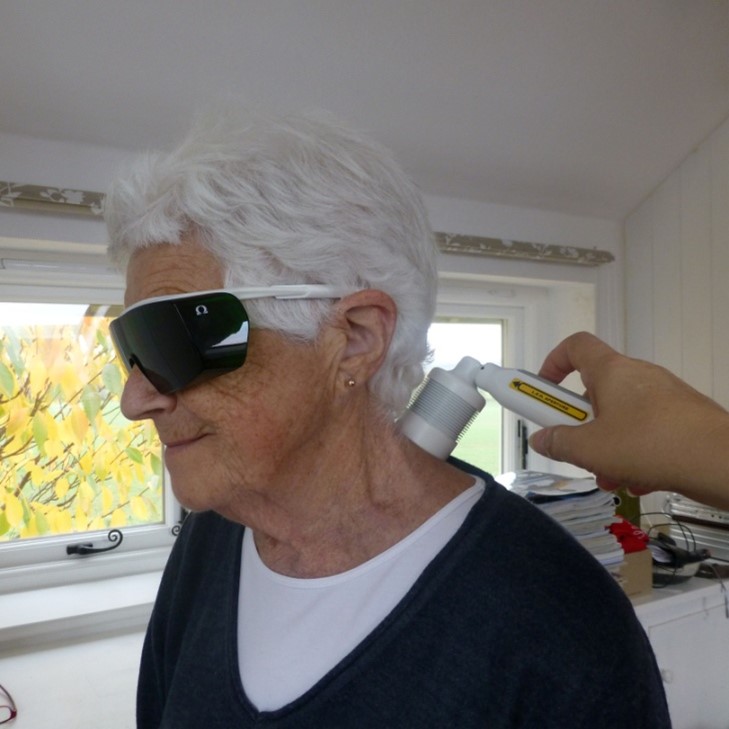
The Xp Laser Systems are LLLT systems that act by photobiomodulation (PBM). This is a form of light therapy that utilises light sources including lasers and light emitting diodes (LEDs) irradiating wavelengths in the visible and near infrared spectrum. It involves the light source being applied to the skin, allowing the light energy (photons) to penetrate tissue where it interacts with chromophores located in cells resulting in photophysical and photochemical changes that lead to alterations at the molecular, cellular and tissular levels of the body. It is a non-thermal, non-ionising process.
It is also therefore very safe with research concluding that the risk of adverse effect arising from the proper use of low level lasers such as the Omega Laser Systems is extremely low ‑ much lower than those of alternative therapies such as pharmacotherapy, including NSAIDs.
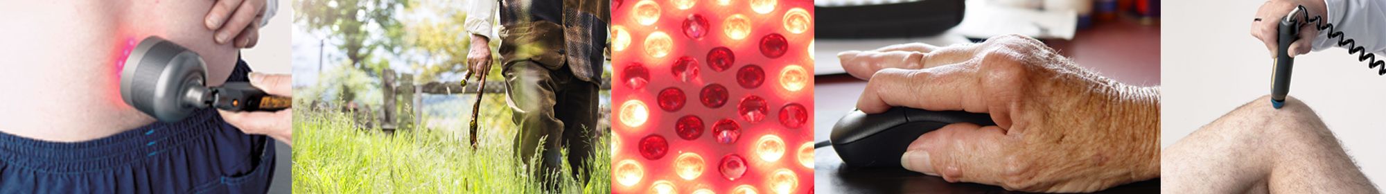
LLLT is commonly used for wound healing and pain relief, being particularly valued in cases where healing processes are otherwise compromised such as with diabetic patients and where pain is persistent, as with arthritic patients.
Rheumatoid Arthritis Pain
Reduction of pain from rheumatoid arthritis is supported by evidence from individual studies as well as in-vitro and non-clinical data, and by the pooled evidence of systematic reviews and meta-analyses, with a moderate short to medium term beneficial effect demonstrated.
Although LLLT appears to modulate pain in more than one way (see below), Lopes-Martin (2007) stated that the overriding biological process for pain relief from LLLT in arthritis is most likely to be its anti-inflammatory action, probably by inhibition of the COX-2 process. The effects seemingly similar to that of Non Steroidal Anti-Inflammatory Drugs.
With respect to LLLT’s ability to modulate inflammatory response, of the 82 laboratory trials and 11 randomised controlled clinical trials identified by systematic search, 71 laboratory trials (87%) provided positive outcomes for one or more parameters, and 7 clinical trials (64%) reported positive results for the reduction of PGE2 levels, reduction of oedema, inflammatory cell infiltration, and reduction of ESR levels (Lopes-Martins et al. 2007). Thus, while there is a degree of variability in reported results, the balance of evidence clearly supports anti-inflammatory actions of LLLT.
There is also strong evidence that this effect is dose-dependent and locally observed in irradiated tissue, and that in cases where no effect was reported, this could be related to inappropriate doses.
Pain Relief Mechanisms
The 2015 Cotler review of current knowledge on LLLT and pain concluded that LLLT appears to modulate pain in more than one way. The peripheral nerve endings of nociceptors, consisting of the thinly myelinated A∂ and unmyelinated, slow-conducting C fibres, lie within the epidermis: this complex network transduces noxious stimuli into action potentials. These nerve endings are very superficial in nature and thus are easily within the penetration depths of the wavelengths used in LLLT but the effects move to nerves in subcutaneous tissues, sympathetic ganglia, and the neuromuscular junctions within muscles and nerve trunks. LLLT applied with an appropriate level of intensity causes an inhibition of action potentials. (Vinck et al. 2005, Chow et al. 2011, Cotler et al. 2015).
It has also been shown that LLLT at the correct dose decreases mitochondrial membrane potential (MMP) in dorsal root ganglion neurons and that ATP production is then reduced. The most immediate effect of nociceptor blockade is pain relief which occurs in a few minutes and has been shown by the timed onset of a conduction blockade in somatosensory-evoked potentials (Hamblin 2017). This inhibition of peripheral sensitization not only lowers the activation threshold of nerves but also decreases the release of pro inflammatory neuropeptides (i.e. substance P and calcitonin gene-related peptide (CGRP)). In persistent pain disorders this reduction of tonic input to activated nociceptors and their synaptic connections, leads to a long-term down-regulation of second-order neurones.
Thus LLLT can have short, medium and long term effects on pain control (Cotler et al. 2015).
Diabetic Ulcers
The clinical evidence from individual studies on LLLT for wound healing in the challenging situation where diabetic ulcers have developed is supportive of benefit from the treatment across in-vitro and non-clinical data, and by the pooled evidence of meta-analyses. The clinical studies demonstrate a moderate beneficial effect on diabetic ulcers over both the short and longer term.
Consistent with our customer feedback, LLLT was also reported in studies to be a pain-free, cost-effective treatment without adverse effects and indeed very few reports of complications were found in the relevant studies. In relation to the state-of-the-art alternatives, it offers a non-invasive method of treatment with a risk to the patient that is very significantly lower than that of any alternative therapy.
Despite considerable variation in treatment regimens and calls for more well-designed trials to be undertaken, all 22 studies on LLLT for diabetic ulcer treatment systematically identified for review reported improved wound healing in terms of either a reduction in ulcer area (cm2) from baseline or between treatment groups, the percentage reduction in ulcer size, or in the proportion (%) of patients with completely or partially healed ulcers.
Wound Healing Action
Wound healing was one of the first applications of LLLT: the therapy can affect all three phases of wound healing: the inflammatory phase, in which immune cells migrate to the wound, the proliferative phase, which results in increased production of fibroblasts and macrophages, and the remodelling phase, in which collagen deposition occurs at the wound site and the extra-cellular matrix is rebuilt.
LLLT is understood to promote wound healing by inducing the local release of cytokines, chemokines, and other biological response modifiers that reduce the time required for wound closure, and increase the mean breaking strength of the wound. This result is achieved by increasing the production and activity of fibroblasts and macrophages, improving the mobility of leukocytes, promoting collagen formation, and inducing neovascularisation (Chung et al. 2012, Hamblin 2017).
In-vitro studies using Omega Laser equipment demonstrate how light irradiation affects macrophage function to either stimulate or inhibit fibroblast proliferation. The in-vitro studies also illustrate that the results obtained are dependent upon the light wavelength, pulse frequency and energy density used, and consequently the importance of determining appropriate irradiation conditions.
Non-clinical studies have shown that light irradiation can stimulate mast cell degranulation which may influence tissue repair and also how LLLT can ameliorate the effect of dermal necrosis following X-irradiation.
After having months of treatment for various leg ulcers from my doctors and nurses, they were not responding. I was referred to Ashdown Clinic, where I received laser treatment. This was successful, and the ulcers were completely healed.
Female patient in nursing / residential home with 23 years leg ulceration. Previously treated using Florozene and Alginate dressings.
Dose: 8 J/cm2 per treatment (2 minutes per point)
Wavelength(s): Multiple wavelengths
Pulsing: mixed low, medium and high frequencies
Probe: 46 Diode Cluster Probe
Frequency/intervals: Once every 2 weeks
Patient reported being pain-free relatively early on in treatment but complete healing of chronic leg ulceration took a total of 9 months (including a 4 month break due to unrelated illness). Quickly able to socialise, no pain and completely healed.

Contact us
Contact Omega to discuss the application of laser in your practice.
Call +44 (0) 1787 477551 or email info@omegalaser.co.uk
